충수 게실염
Appendiceal Diverticulitis
Article information
Abstract
복통과 우하복부 압통, 반발통을 보인 64세 남자에서 충수벽 외부로 돌출되는 낭, 낭 안의 고에코 분석, 게실과 연결된 충수벽의 비후, 게실 주위 염증성 지방침윤으로 인한 고에코 병변 등 충수 게실염의 전형적인 초음파 소견을 관찰할 수 있었고, 수술과 병리조직 결과를 통해 확인할 수 있었다
Trans Abstract
Appendiceal diverticulitis is an uncommon cause of right lower quadrant abdominal pain. Mostly, it is considered as acute appendicitis because the clinical presentation of the appendiceal diverticulitis is similar to acute appendicitis; therefore it is diagnosed by appendectomy and a pathologic specimen. The patients with appendiceal diverticulitis have higher rates of perforation. Also, they need longer operation times and longer postoperative hospital stays. They are relatively old and have a longer duration of symptoms. We report a 64-year-old male case of appendiceal diverticulitis with perforation. It was diagnosed by ultrasound and confirmed by a surgical specimen. Preoperative diagnosis of appendiceal diverticulitis is not impossible using high resolution ultrasound equipment. Although there are limits to diagnosing appendiceal diverticulitis by ultrasound, we must give particular attention to a clinical setting which makes it possible.
서 론
충수 게실염은 우하복부 통증을 유발하는 드문 질환이다. 일반적인 급성 충수염과 임상 양상이 유사하므로, 대부분 급성 충수염으로 진단되어 수술이 시행되며, 최종적으로 병리 결과를 통해 확인되는 경우가 흔하다. 충수 게실염은 일반적인 급성 충수염에 비해 천공 등 합병증이 동반되는 경우가 많으므로, 이와 연관된 임상 양상과 초음파 소견을 숙지하여야 조속한 치료와 양호한 경과를 얻을 수 있다.
증 례
64세 남자 환자가 급격히 심해진 복통으로 내원하였다. 환자는 10일 전 본원에서 특별한 증상 없이 건강검진으로 대장내시경검사를 받았고, 당시 상행결장에 약 8 mm 크기의 유경성 용종이 한 개 발견되어 올가미를 이용한 용종절제술을 시행 받았다. 이후 특별한 문제없이 지내다, 내원 2일 전 과음한 후 복통과 설사가 발생하였고, 내원 전날 저녁부터는 심한 복통으로 잠을 이룰 수가 없었다. 환자는 특별한 과거 병력은 없었으며, 1주에 3-4회 음주하였고 하루에 한 갑 흡연하였다. 내원 당시 환자는 급성 병색을 보였고, 신체검사에서 장음은 약간 증가되어 있었다. 우하복부에는 압통과 반발통을 보였고, 체온은 36.5°C로 정상이었다. 초음파(Samsung Medison H60, Samsung Medison Co. Ltd., Seongnam, Korea) 검사를 시행하였고, 충수의 확장(maximal outer diameter 9.7 mm)과 충수벽의 비후가 관찰되었다. 충수 체부에서 내부 고에코 부분을 가진 외부로 돌출된 낭이 관찰되었고, 이와 연결된 충수 체부의 벽비후와 돌출된 낭 주위로 염증성 지방침윤으로 여겨지는 고에코 병변이 관찰되었다(Fig. 1). 이는 대장 게실염에서 보이는 초음파 소견과 유사하며, 전형적인 충수 게실염의 초음파 소견으로 볼 수 있다. 환자는 당일 복강경하 충수절제술을 시행 받았고 4일 후 합병증 없이 퇴원하였다.

The ultrasonographic findings of appendiceal diverticulitis. (A) The oblique scan using a low frequency convex probe at the right lower quadrant of the abdomen shows the cecum (asterisk) and the appendiceal base (arrow). (B) Using a high frequency linear probe, the scan shows mild wall thickening at the appendiceal base (arrow). (C) Using a high frequency linear probe, shows moderate wall thickening at the appendiceal body and hyperechoic fat infiltration. Maximum outer diameter (between plus-markers) of the appendiceal body is 9.7 mm. (D) The outpouching sac from the appendiceal body corresponds to the appendiceal diverticulum (asterisk).
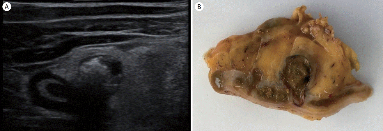
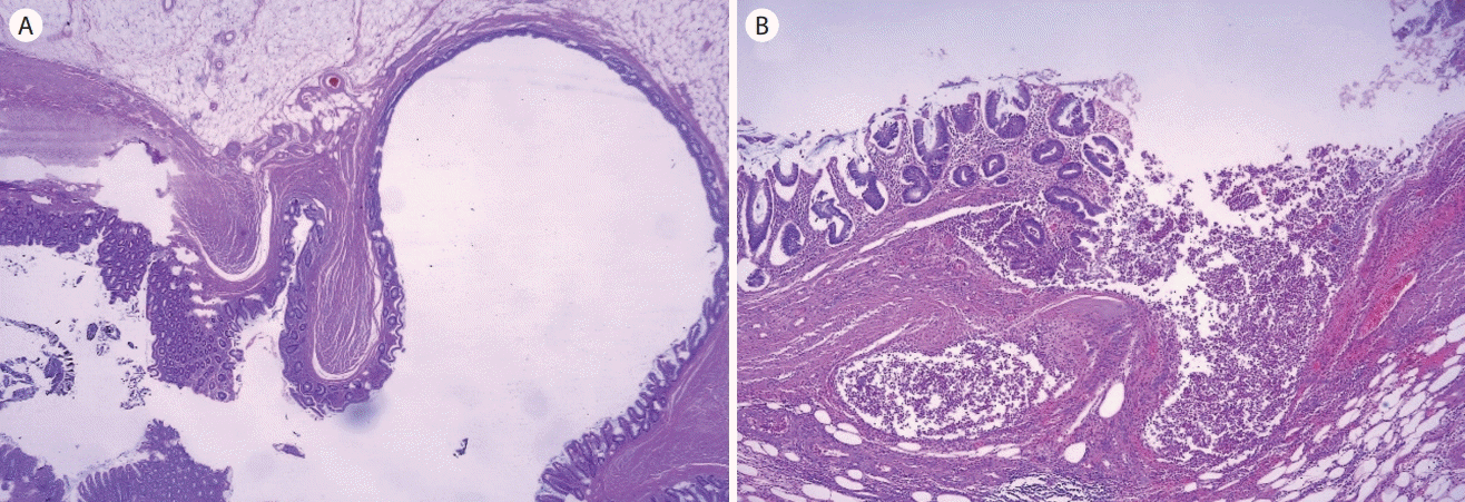
절제된 검체에서 분변으로 채워진 충수 게실과 이와 연결된 충수벽의 비후, 그리고 충수 게실 주위 지방 침윤 소견이 있었고(Fig. 2), 현미경검사에서 충수 주위 염증 세포의 침윤 및 천공이 확인되어 “천공과 충 수 주위 농양을 동반한 충 수 게실염”으로 최종 진단되었다(Fig. 3).

The ultrasonographic finding and pathologic specimen of appendiceal diverticulitis. (A) The ultrasonogram shows the outpouching sac with internal hyperechoic foci, thickened appendiceal walls, and hyperechoic fat infiltration around the sac. (B) The pathologic specimen shows impacted fecal material within the appendiceal diverticulum, thickened appendiceal walls, and peridiverticular fat infiltration. It also shows a tiny diverticulum at the proximal site.
고 찰
충수 게실염은 우하복부 통증을 유발하는 드문 질환이다. 충수절제술 후 발견되는 충수 게실염의 빈도는 0.004%에서 2.1%까지 보고되었고, 통상의 부검을 통해서는 0.2%에서 0.6%까지 보고되었다[1,2]. 충수 게실염 환자의 임상 양상은 급성 충수염 환자의 임상 양상과 유사하며, 충수 게실염은 드문 질환이라 간과되어, 수술 전에는 급성 충수염으로 진단되는 경우가 대부분이다. 충수 게실염과 일반적인 급성 충수염의 차이점을 보면, 충수 게실염에서 천공의 발생이 높고, 그로 인해 더 긴 수술 시간과 수술 후 회복 시간을 가진다. 저자에 따라 충수 게실염의 천공발생률이 4-6배 높다고 보고하였다[3,4]. 또한 충수 게실염 환자는 급성 충수염 환자에 비해 나이가 많고, 증상이 발생하고 수술하기까지 걸리는 시간도 더 길다[3,4].
충수의 게실성 질환은 다음과 같이 총 4가지 형태로 분류된다[5]: 1) 염증이 동반되지 않은 충수 게실(appendiceal diverticula without inflammation), 2) 정상 충수 게실을 가진 급성 충수염(acute appendicitis with diverticula), 3) 급성 충수염과 급성 충수 게실염을 함께 보이는 경우(acute appendiceal diverticulitis with acute appendicitis), 4) 충수 게실에만 염증이 국한된 급성 충수 게실염(acute appendiceal diverticulitis). 본 증례는 세 번째 경우에 해당한다.
충수 게실염의 초음파 소견은 대장 게실염의 초음파 소견과 유사하다. 충수벽 외부로 돌출되는 낭을 가지며, 낭 안에는 고에코의 분석이 관찰될 수 있다. 또한 게실과 연결된 충수벽의 비후를 보이며, 게실 주위로는 염증성 지방침윤으로 인해 고에코의 병변을 보인다[3,5]. 이번 증례에서도 이러한 초음파 소견을 모두 관찰할 수 있었다.
전형적인 급성 화농성 충수염과 충수 게실염의 초음파 감별점을 보고한 논문에 따르면, 전자는 고에코의 두꺼워진 충수벽(점막층 및 점막하층의 염증성 변화)과 내부 무에코(염증에 의한 액체 성분)를 보이는 반면, 후자는 충수 게실 안에 공기에 의한 고에코 부분이 나타난다고 하였다[5]. 본 증례에서도 초음파 검사에서 충수 게실 내 고에코 소견이 보였으나, 공기에 의한 것은 아니었고 게실 내 분변 매복에 의한 것이었다.
증상이 있는 충수 게실염은 천공 등 합병증 발생 빈도가 높기 때문에 신속한 수술적 치료가 이루어져야 한다. 무증상의 충수 게실에 대해서는 아직 이론이 있으나, 충수 게실염 발생 가능성이 높고 발생시 천공 등 합병증 동반이 많으며, 악성 질환의 동반 가능성도 있어 예방적 수술이 권고되고 있다.
초음파 장비가 향상되면서 고해상도 초음파를 통해 수술 전 충수 게실염을 진단한 증례가 늘어나고 있다[3,5]. 충수 게실염의 임상 양상과 초음파 소견을 숙지하여, 수술 전 초음파검사에서 충수 게실염 진단이 간과되지 않도록 하는 것이 중요하겠다.
