수술 전 초음파검사로 진단한 충수 게실염
Preoperative Diagnosis of Diverticulitis of the Vermiform Appendix by Ultrasonography
Article information
Abstract
충수 게실염은 드문 질환으로 보고되고 있고 임상적으로 급성 충수염과 구분이 어려워 수술 전 진단이 어려운 현실이다. 충수 게실염은 급성 충수염에 비해서 발생 연령이 높고 증상이 심하지 않고 증상의 지속시간이 긴 특징이 있으며 천공률이 높아서 조기 발견 및 치료가 중요하다. 또한 우하복부 통증의 병력이 동반되는 경우가 있어서 반복적인 우하복부 통증 환자에서 복부 초음파검사를 하여 충수 게실을 확인하는 것은 의의가 있을 것이다. 최근에는 고해상도 초음파 기계가 보급되면서 우하복부 통증 환자에서 이전에 비해 충수 게실염의 발견이 증가되고 있는 추세이며 임상 양상 및 특징적인 초음파 소견을 숙지하는 것이 필요하겠다.
Trans Abstract
Diverticulitis of the vermiform appendix is an uncommon condition, mimicking appendicitis. Several differences have been observed. Compared with acute appendicitis, diverticulitis of the vermiform appendix has a longer duration of pain; primarily develops in older adults; has a lower frequency of accompanying abdominal pain, nausea and vomiting; has a high risk of perforation. Preoperative diagnosis is rare, but can be achieved by high resolution ultrasonography. So it is meaningful to keep in mind the symptoms and ultrasonographic findings of diverticulitis of the vermiform appendix in patients with lower abdominal pain. Author report a case of ultrasonographic diagnosis of diverticulitis of the vermiform appendix in an adult male.
서 론
충수 게실염은 드문 질환이며 우하복부 통증 환자의 진단적인 검사에서 간과되는 경우가 많다. 임상 양상이 급성 충수염과 비슷하지만 치료와 예후 면에서 약간의 차이가 있으므로 수술 전 진단이 의미가 있는 질환이다.
국내에서는 충수염을 의심하여 수술한 환자에서 충수 게실염을 진단하였던 논문은 있었으나 수술 전에 초음파검사로서 충수 게실염을 진단하였던 예는 거의 없었다. 저자는 우하복부 통증으로 내원한 중년 남성에서 초음파검사로 충수 게실염을 진단하였기에 보고한다.
증 례
57세 남자가 3일간의 우하복부 통증을 주소로 내원하였다. 진찰상 우하복부 압통을 호소하였다. 체온은 36.5도로 정상이었고, 혈액검사에서 WBC 12,500 (poly 76%), Hb 13.4g/dL, platelet 356,000이었다.
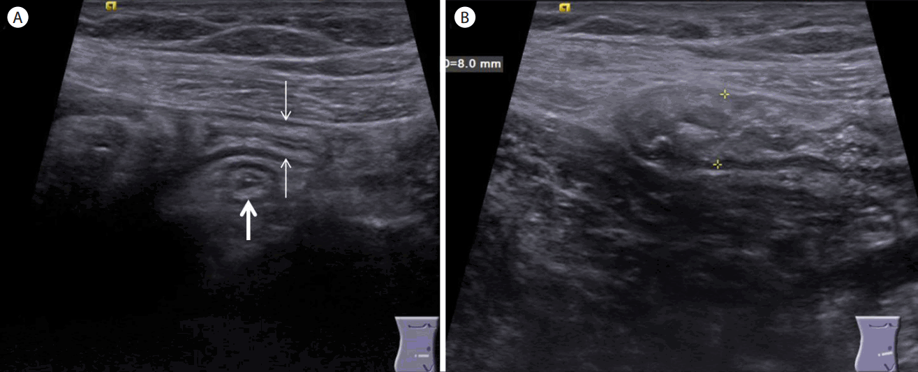
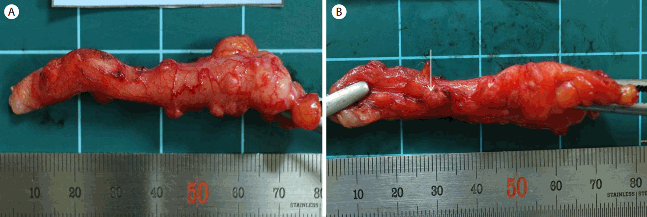
복부 초음파검사(Acuson S1000, Siemens, Germany)에서 우하복부에 탐촉자로 압박이 되지 않는 종대된 충수가 보였고(전후 직경 8 mm; Fig. 1) 충수의 체부와 말단부에 충수벽외로 돌출하는 5 mm 크기의 저에코의 병변과 주위에 고에코의 지방침착이 관찰되었다(Fig. 2). 도플러검사에서 충수벽과 충수벽 외로 돌출하는 충수 게실 주변에 혈류증가가 관찰되어(Fig. 2) 급성 충수염과 충수 게실염이 같이 동반된 증례가 의심되어 일반외과로 전원되어 충수절제술을 시행받았다. 수술 소견은 충수가 8 mm로 종대되어 있고 화농성 염증 소견이 관찰되었다. 충수벽 외로 돌출되는 0.4-0.6 cm 크기의 다발성의 충수 게실이 관찰되었고 게실 중 저명한 염증성 병변이 관찰되었고 천공은 없었다(Fig. 3). 병리학적 소견은 충수벽과 충수 게실벽의 염증세포 침윤과 충수 게실벽 내의 점막하층에 섬유성 변화가 관찰되었다(Fig. 4). 충수 게실벽은 점막층과 점막하층으로 구성되어 있어서 가성 게실이 의심되었다.

(A) Ultrasonogram showed normal long axis view (arrows) and short axis view of the proximal appendix (thick arrow). (B) Ultrasonogram showed and distended body of appendix (calipers, 8 mm,) with thickened appendiceal wall (3.3 mm, calipers not shown, B).

(A) Ultrasonogram showed round hypoechoic outpouching (arrow) at mesoappendiceal margin with peridiverticular hyperechoic fat infiltration near the tip of the appendix (thick arrow). (B) Color doppler ultrasonogram showed increased flow within the diverticular wall.

(A) The resected specimen showed distended appendix with moderate inflammation with engorged vessels. Also multiple diverticula (0.4-0.6 cm in diameter) protruding from the wall of the appendix were seen. Any perforations were not seen. (B) Prominent appendiceal diverticulum with peridiverticular inflammation was seen (arrow).

Histologic features of appendiceal diverticulitis. Note that the wall of the appendiceal diverticulum was composed of mucosa, muscularis mucosa and submucosa which is compatible with pseudodiverticulum. Inflammatory cell infiltration and submucosal fibrosis were noted within the wall of the appendiceal diverticulum (H&E, ×10).
고 찰
충수에 발생하는 게실은 드문 질환으로서 1893년 Kelynack [1]에 의해 처음 기술된 이래 여러 논문에서 발표되어 왔으며 발생 빈도는 0.004% [2]에서 2.1% [3]까지 다양하다.
충수에서 발생하는 게실은 진성 게실 또는 가성 게실로 구분되며 대부분의 경우 후천적으로 발생하는 가성 게실의 형태로 보인다. 진성 게실은 게실 벽이 점막층, 점막하층, 근층으로 구성되어 있으나, 가성 게실은 점막층과 점막하층으로 구성되어 있어서 천공의 가능성이 높다.
충수의 가성 게실 발생 기전은 충수 내강이 막히거나 내강의 압력이 높아지고 충수 근육층의 수축에 의해 근육층 결손부로의 점막층과 점막하층이 탈출됨으로써 발생된다고 보고하였다[4].
가성 게실의 발생 가설에 염증성과 비염증성 가설로 구분해서 설명하고 있다. 염증성 가설은 반복적인 염증과 감염으로 인해 충수벽의 림프조직의 위축이 발생하고 이로 인해 충수벽이 약해지고 얇아짐으로써 발생한다고 한다. 반면 비염증성 가설은 충수 내강의 협착, 충수 분석, 종양 등으로 인해 충수 내강의 압력 증가와 근육층의 수축으로 인해 게실이 발생되는 것으로 알려져 있다[4].
충수 게실염의 원인은 충수 내강의 폐색이 오고 이로 인해 충수 게실벽의 부종 및 출혈을 유발하고 과다한 점액분비로 염증과 천공을 일으키는 것으로 알려져 있지만 충수 내강의 폐색이 모든 예에서 발견되는 것이 아니어서 명확한 이유는 알려져 있지 않다. 충수 게실은 약 60%에서 충수 원위부에서 발생하고 게실의 수는 보통 한 개에서 수개까지 보고되고 있으며[5] 본 증례에서도 체부와 원위부에서 다발성으로 발현되었다.
충수 게실염과 급성 충수염과는 발생 연령에서 차이가 있다. 대부분의 충수 게실증과 충수 게실염은 30대, 40대에 호발하는 것으로 보고되고 있어서 급성 충수염의 호발 연령인 10대, 20대와 차이를 보여준다[5].
충수 게실염의 증상은 우하복부 혹은 배꼽주위 통증으로 나타나지만 복통 발현 후 내원까지의 시간 경과가 급성 충수염이 보통 24시간 이내인 것에 비해 60%에서 48시간 이후에 내원한 것으로 보고되고 있고[6] 이는 충수염에 비해서 통증이 경미하거나 모호하기 때문으로 생각된다.
충수 게실염에서의 통증은 종종 재발되는 양상을 보여서 경우에 따라서 내원 전 수주 또는 수개월 동안 복부 통증을 경험한 경우가 있다. 따라서 반복되는 우하복부 통증을 호소하는 환자에서 복부 초음파검사로 충수 게실의 유무를 확인하는 것은 의미가 있다.
급성 충수염의 천공률은 15%에서 30%까지 보고되고 있는 반면 충수 게실염의 경우 27-66%로 보고하고 있다[4]. 이는 충수 게실염의 경우 대부분이 가성 게실로서 근육층이 없고 게실이 주로 장간막 경계부에 위치해서 체간통 발현이 잘 나타나지 않기 때문으로 알려져 있다.
충수의 게실성 질환은 4가지 형태로 구분하여 기술한 바 있는데[7] 이는 1) 염증이 없는 충수 게실(appendiceal diverticula without inflammation), 2) 급성 충수염에 동반된 정상 충수 게실(acute appendicitis with appendiceal diverticula), 3) 급성 충수염과 동반된 충수 게실염(acute appendiceal diverticulitis with acute appendicitis), 4) 충수 게실염(acute appendiceal diverticulitis)의 형태로 구분되며 이 중에서 네 번째의 경우가 진정한 충수 게실염에 해당하나 본 증례는 충수염과 충수 게실염이 동반된 경우였다.
충수 게실염의 대부분은 급성 충수염이 의심되어 수술한 환자에서 수술 후 충수 게실염으로 진단되었던 경우이다. 한 연구에 의하면 급성 충수염으로 수술받았던 환자들 중 2%는 충수 게실염이었다는 보고를 한 바 있다[8]. 이는 충수 게실염과 급성 충수염의 병력과 임상 양상이 비슷하기 때문에 수술 전 정확한 진단이 어렵기 때문이다.
충수 게실염의 수술 전 영상 의학적 진단에 대한 보고는 흔치 않다. Place 등[5]은 충수 게실염의 복부 전산화단층촬영(computed tomography, CT) 소견에 대해 기술하였는데 맹장 게실염과 감별이 불가능하였으며 이는 CT의 해상도 때문에 작은 충수 게실을 간과하였기 때문이라고 하였다. 최근에는 고해상도 초음파검사로서 충수 게실염을 진단한 외국의 보고는 있었으나 국내에서는 거의 없는 실정이다.
충수 게실염의 초음파 소견은 충수 벽 외로 돌출하는 원형의 저에코성 게실과 그 주변의 고에코 지방 침착 소견이 있다. 색 도플러 검사에서 충수 게실과 주위로 혈류증가 소견을 관찰할 수 있다. 주위에 있는 맹장과 말단 회장벽 비후 소견이 관찰되는 경우도 있으며 천공이 되면 게실의 연속성이 파괴되고 체액 저류가 관찰된다.
Kubota 등[9]의 연구에 의하면 전형적인 급성 화농성 충수염에서는 종대된 충수 내강에 점액과 농에 의한 무에코가 관찰되는 반면 충수 게실염에서는 충수 게실이 위치한 충수 벽의 비후와 충수 벽 외로 돌출하는 저에코의 충수 게실과 동반하여 충수 내강에는 정상적인 공기 음영이 관찰된다고 하였다. 그 이유는 급성 충수염은 충수 내강의 폐쇄로 인한 질환인 반면 충수 게실염은 충수 게실이 위치한 충수 벽의 염증성 변화이기 때문이라고 하였다.
국내에서는 36예의 충수 게실염에 대한 보고[10]가 있었으나 이는 충수염을 의심하여 수술 시행 후 충수 게실염으로 진단되었던 경우였다. 반면에 본 증례는 우하복부 통증을 주소로 내원하여 수술 전에 초음파검사로서 충수 게실염을 진단받고 수술 후 확진되었다는 점에서 의미가 있겠다.
충수 게실염의 치료는 가능하면 즉각적인 수술적 치료가 요구된다. 무증상 충수 게실이 발견되는 경우 염증, 출혈 및 천공 등의 위험성이 있어 수술적인 치료를 권유한다[7].