경부 림프절의 초음파 진단

Ultrasonographic Findings of Cervical Lymph Nodes

Article information

Clin Ultrasound. 2023;8(2):48-52
Publication date (electronic) : 2023 November 30
doi : https://doi.org/10.18525/cu.2023.8.2.48
Division of Endocrinology and Metabolism, Department of Internal Medicine, Yeungnam University Medical Center, Yeungnam University College of Medicine, Daegu, Korea
정승민
영남대학교 의과대학 영남대학교병원 내분비내과
Address for Correspondence: Seung Min Chung, M.D., Ph.D. Division of Endocrinology and Metabolism, Department of Internal Medicine, Yeungnam University Medical Center, Yeungnam University College of Medicine, 170 Hyunchung-ro, Nam-gu, Daegu 42415, Korea E-mail: smchung@ynu.ac.kr
Received 2023 September 20; Revised 2023 October 15; Accepted 2023 October 25.

Trans Abstract

Ultrasonography is an accurate and cost-effective diagnostic tool for cervical lymph node assessment. This article briefly reviews the key grayscale (size, shape, echogenicity, fatty hilum, and calcification) and color Doppler ultrasound findings to help distinguish benign and malignant lymph nodes.

서 론

경부 림프절의 초음파 진단은 갑상선 결절이 있거나 새로 갑상선암이 진단된 환자의 림프절 상태를 평가하고, 치료받은 갑상선암 환자의 재발성 질환을 발견하는데 중요한 역할을 한다. 초음파를 통해 림프절의 위치 파악은 물론, 그 종대가 병적인 것인지에 대한 판정을 하게 된다. 갑상선암 환자의 술 전 경부 림프절 초음파는 갑상선 유두암 환자의 최대 30%에서 임상적으로 만져지지 않는 전이성 림프절을 발견할 수 있고, 이로 인해 최대 40%의 환자에서 수술 방법이 변경될 수 있다[1,2]. 즉, 술 전 부적절한 림프절 평가는 암의 잔존 혹은 재발을 초래하므로 림프절 전이의 가능성을 명심하고 전 경부를 면밀히 검사 하여야 한다. 본론에서는 양성 및 전이성 경부 림프절 소견에 대해 살펴보고자 한다.

본 론

지금까지 많은 연구자들에 의해 림프절의 크기, 내부 특징, 경계 특징, 도플러 소견 등을 참고로 양성과 전이성 림프절의 감별을 위한 진단 기준이 검토되어 왔다[3-6]. 2021년 대한갑상선영상의학회에서 개정 발표한 갑상선 결절의 악성 위험도 분류체계(Korean Thyroid Imaging Reporting and Data System, K-TIRADS) 권고안에 따르면 경부 림프절의 전이 위험은 초음파 소견에 따라 다음과 같이 세 분류로 나눌 수 있다[7].

  • - 전이 의심(Suspicious): Cystic change, echogenic foci (calcification), cortical hyperechogenicity (focal/diffuse), abnormal vascularity (peripheral/diffuse)

  • - 미확정(Indeterminate): Loss of echogenic hilum and hilar vascularity

  • - 양성(Probably benign): Echogenic hilum, hilar vascularity

양성 림프절

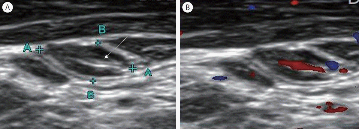
양성 림프절은 균일한 저에코의 편평한 타원형(단축/장축<0.5) 모양을 띄며 경계가 명확하다[8]. 림프절의 크기는 대개 단축에서는 5 mm를 넘지 않고 장축에서는 5–10 mm 전후이나 2–3 cm 길이의 림프절도 정상적인 성인과 어린이에게서 발견되므로 크기 단독은 양성과 악성을 감별하기에 좋은 기준이 아니다[9-11]. 림프절 변연부에서 중심부를 향해 띠모양으로 뻗어 있는 고에코 음영의 문(central echogenic hilus)은 지방문(fatty hilum) 의 존재를 시사하며 이는 민감도 100%로 양성 림프절로 간주할 수 있는 소견이다[6,12]. 도플러 소견으로는 림프절 문으로만 분포하는 혈류신호가 관찰되는데 림프절의 위치, 성별, 연령에 따라 혈류신호가 전혀 관찰되지 않는 경우도 있다(Fig. 1) [13].

Figure 1.

Transverse ultrasound image of benign lymph nodes. (A) Echogenic hilum (arrow). (B) Hilar vascularity.

갑상선 분화암의 림프절 전이

갑상선 유두암 및 갑상선 여포암은 약 20–50%의 빈도로 림프절 전이를 수반하는 질환이다[14]. 갑상선 분화암에서 림프절 전이를 시사하는 소견으로는 림프절의 종대, 림프절 문의 소실, 원형 모양, 낭성 변화, 점상 고에코 소견, 림프절 피질 고에코 소견, 비정상 혈류(림프절 변연부 혹은 림프절 문과 변연부 모두에 혈류 신호 관찰)가 있다[12,14]. 대개 악성 림프절은 인접 근육과 비교 시 저에코인 반면 갑상선 유두암의 전이 림프절은 고에코를 나타내는 경우가 많은데 이는 갑상선글로불린(thyroglobulin)의 침적과 관련이 있다[15]. 석회화는 림프절의 변연에 점상으로 혹은 중심에 모여 있는 양상을 나타내고 후방음영(acoustic shadowing)을 동반하는 경우도 있다[15]. 낭성 변화, 점상 고에코 소견은 특이도 100%로 림프절 전이를 강하게 의심할 수 있는 소견이고, 림프절 변연부의 혈류 증가는 림프절 전이 예측에 있어 민감도(86%)와 특이도(82%)의 균형이 가장 잘 잡힌 소견이다(Fig. 2) [6].

Figure 2.

Transverse ultrasound image of metastatic lymph nodes of papillary thyroid carcinoma. (A) Diffuse hyperechogenicity without an echogenic hilum (washed thyroglobulin level was 22.70 ng/mL). (B) Intranodal cystic change. (C) Punctate echogenic foci (microcalcifications) and macrocalcifications with posterior acoustic shadowing. (D) Peripheral and central increased vascularization.

갑상선 수질암의 림프절 전이

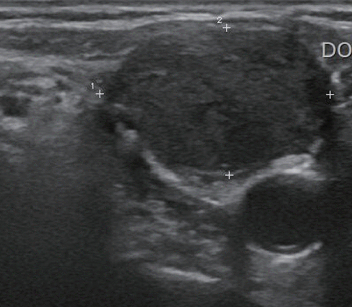
갑상선 수질암은 약 40–80%의 빈도로 림프절 전이를 수반하는 질환으로 갑상선 분화암에 비하여 림프절 전이가 흔하다[16]. 갑상선 수질암과 갑상선 유두암의 초음파 소견을 비교하였을 시 갑상선 수질암에서 난형-원형 모양(ovoid to round shape)이 더 흔한 것 외에는 특별한 차이가 없었다[17]. 갑상선 수질암과 갑상선 유두암의 전이성 림프절의 초음파 소견을 비교한 후향적 연구에 따르면 갑상선 수질암의 전이성 림프절이 크기가 더 크고, 림프절 문 소실이 더 흔하며, 석회화가 적은 경향이 있었다[18]. 갑상선 수질암을 시사하는 전형적인 초음파 소견은 없으나 초음파 상 고형종이며 악성 의심도가 높은 결절을 시사할 시에는 갑상선 수질암의 가능성을 염두에 두고 선별적으로 혈청 칼시토닌을 검사하는 것이 좋겠다(Fig. 3) [19].

Figure 3.

Transverse ultrasound image of a metastatic lymph node of medullary thyroid carcinoma. The lymph node was located in the right level III, measured 2.10 × 1.34 cm, and was hypoechoic without an echogenic hilum. The serum calcitonin level was >740 pg/mL.

림프종

경부 악성 림프절은 림프종 침범이 흔히 일어나는 부위로 림프절종의 다른 원인과 치료 방법이 다르기 때문에 정확한 진단이 필수적이다. 림프절 크기는 대개 단축에서 10 mm 이상으로 커져 있고 둥근 모양, 저에코 음영에 림프절 문이 소실되어 있다[20]. 림프절 내 낭성 변화는 방사선 치료를 하였거나 진행성 질환(advanced disease)이 아닌 이상은 거의 발견되지 않고, 도플러 소견으로는 62–90%에서 림프절 문과 변연부에 함께 혈류가 증가한다[20]. 림프종성 림프절에서는 가성낭성(pseudocystic) 양상에 더불어 후방음향의 강화로 낭성 림프절로 오인할 수 있는데, 고해상도 초음파를 사용하여 림프종성 림프절의 특징적인 미세결절 망상 모양(micronodular reticular echopattern)을 확인하는 것이 도움이 된다(Fig. 4) [8,12].

Figure 4.

Transverse ultrasound image of non-Hodgkin’s lymphoma. The lymph node was located in the left supraclavicular area, measured 3.56 × 2.24 cm, and showed intranodal reticulation.

원격 림프절 전이

경부의 악성 림프절은 대부분 두경부 원발성 종양의 전이 혹은 림프종이지만, 때때로 쇄골 하 부위의 원발암이 원격 전이되기도 한다. 일반적으로 목 상부 2/3의 전이성 림프절은 머리와 목 기원의 원발성 종양에서 유래하고, 목 하부 1/3과 쇄골 상 전이성 림프절은 유방, 폐 등의 전이성 질환의 가능성이 높으나 이런 원격 전이는 목 상부에서 관찰되기도 한다(Fig. 5) [21]. 초음파와 미세침 흡인 세포검사는 비용이 저렴하고 진단 정확도가 높기 때문에 경부 림프절의 종대, 원형 모양, 저에코 소견, 림프절 문의 소실, 비정상 혈류 소견 등이 있을 시 원격전이를 의심하고 시행하는 것이 권장되며, 면역조직화학검사를 통해 보다 정확한 진단이 가능하다.

Figure 5.

Transverse ultrasound image of metastatic lymph nodes of lung cancer. (A) Hypoechoic, cortical hyperechogenicity without an echogenic hilum. (B) Cystic necrosis.

결 론

경부 림프절의 진단을 위해서는 림프절의 크기, 모양, 경계, 에코 음영, 림프절 문, 혈류 양상 소견을 종합하여 판단해야 한다. 갑상선 초음파 상 갑상선암 의심 소견이 있을 시에는 림프절 전이의 가능성을 명심하고 전 경부를 면밀히 검사하여야 한다. 반대로 악성이 의심되는 경부 림프절을 발견했을 시에는 갑상선의 면밀한 관찰을 포함하여 림프종, 기타 원발암의 전이 등을 염두에 두고 접근해야 하겠다.
References

1. Kouvaraki MA, Shapiro SE, Fornage BD, et al. Role of preoperative ultrasonography in the surgical management of patients with thyroid cancer. Surgery 2003;134:946–954. discussion 954-955.
2. Stulak JM, Grant CS, Farley DR, et al. Value of preoperative ultrasonography in the surgical management of initial and reoperative papillary thyroid cancer. Arch Surg 2006;141:489–494. discussion 494-496.
3. Chung SR, Baek JH, Rho YH, et al. Sonographic diagnosis of cervical lymph node metastasis in patients with thyroid cancer and comparison of European and Korean guidelines for stratifying the risk of malignant lymph node. Korean J Radiol 2022;23:1102–1111.
4. Lee JY, Yoo RE, Rhim JH, et al. Validation of ultrasound risk stratification systems for cervical lymph node metastasis in patients with thyroid cancer. Cancers (Basel) 2022;14:2106.
5. Shin YG, Kang H, Joh YD, Jeong KS, Kim BS. Ultrasonographic features of metastatic lymph nodes in papillary thyroid microcarcinomas and macrocarcinomas. J Korean Soc Radiol 2015;72:92–100.
6. Leboulleux S, Girard E, Rose M, et al. Ultrasound criteria of malignancy for cervical lymph nodes in patients followed up for differentiated thyroid cancer. J Clin Endocrinol Metab 2007;92:3590–3594.
7. Ha EJ, Chung SR, Na DG, et al. 2021 Korean thyroid imaging reporting and data system and imaging-based management of thyroid nodules: Korean society of thyroid radiology consensus statement and recommendations. Korean J Radiol 2021;22:2094–2123.
8. Dudea SM, Lenghel M, Botar-Jid C, Vasilescu D, Duma M. Ultrasonography of superficial lymph nodes: benign vs. malignant. Med Ultrason 2012;14:294–306.
9. Iwata M, Kasagi K. Ultrasonographic diagnosis of thyroid and neck 2nd edth ed. Seoul: Medianbook; 2007.
10. Lee JC. Ultrasound of neck mass. J Clinical Otolaryngol 2007;18:152–160.
11. Jeon YS. Ultrasonographic evaluation of cervical lymph nodes. J Surg Ultrasound 2023;10:1–7.
12. Prativadi R, Dahiya N, Kamaya A, Bhatt S. Chapter 5 ultrasound characteristics of benign vs Malignant cervical lymph nodes. Semin Ultrasound CT MR 2017;38:506–515.
13. Ying M, Ahuja A, Brook F, Metreweli C. Power Doppler sonography of normal cervical lymph nodes. J Ultrasound Med 2000;19:511–517.
14. Haugen BR, Alexander EK, Bible KC, et al. 2015 American thyroid association management guidelines for adult patients with thyroid nodules and differentiated thyroid cancer: The American thyroid association guidelines task force on thyroid nodules and differentiated thyroid cancer. Thyroid 2016;26:1–133.
15. Ahuja A, Ying M. Sonography of neck lymph nodes. Part II: abnormal lymph nodes. Clin Radiol 2003;58:359–366.
16. Wells SA Jr, Asa SL, Dralle H, et al. Revised American Thyroid Association guidelines for the management of medullary thyroid carcinoma. Thyroid 2015;25:567–610.
17. Kim SH, Kim BS, Jung SL, et al. Ultrasonographic findings of medullary thyroid carcinoma: a comparison with papillary thyroid carcinoma. Korean J Radiol 2009;10:101–105.
18. Ni X, Xu S, Zhan W, Zhou W. Ultrasonographic features of cervical lymph node metastases from medullary thyroid cancer: a retrospective study. BMC Med Imaging 2022;22:151.
19. Park MJ, Choi YS, Song HS, Kim BS. The relationship between ultrasonographic features and clinical characteristics of medullary thyroid carcinoma. Clin Ultrasound 2018;3:8–14.
20. Ahuja AT, Ying M, Ho SY, et al. Ultrasound of malignant cervical lymph nodes. Cancer Imaging 2008;8:48–56.
21. López F, Rodrigo JP, Silver CE, et al. Cervical lymph node metastases from remote primary tumor sites. Head Neck 2016;38(Suppl 1):E2374–E2385.

Article information Continued

Figure 1.

Transverse ultrasound image of benign lymph nodes. (A) Echogenic hilum (arrow). (B) Hilar vascularity.

Figure 2.

Transverse ultrasound image of metastatic lymph nodes of papillary thyroid carcinoma. (A) Diffuse hyperechogenicity without an echogenic hilum (washed thyroglobulin level was 22.70 ng/mL). (B) Intranodal cystic change. (C) Punctate echogenic foci (microcalcifications) and macrocalcifications with posterior acoustic shadowing. (D) Peripheral and central increased vascularization.

Figure 3.

Transverse ultrasound image of a metastatic lymph node of medullary thyroid carcinoma. The lymph node was located in the right level III, measured 2.10 × 1.34 cm, and was hypoechoic without an echogenic hilum. The serum calcitonin level was >740 pg/mL.

Figure 4.

Transverse ultrasound image of non-Hodgkin’s lymphoma. The lymph node was located in the left supraclavicular area, measured 3.56 × 2.24 cm, and showed intranodal reticulation.

Figure 5.

Transverse ultrasound image of metastatic lymph nodes of lung cancer. (A) Hypoechoic, cortical hyperechogenicity without an echogenic hilum. (B) Cystic necrosis.