화농성 비장농양과 감별이 어려운 속발성 비장결핵 의심 증례
Suspected Case of Secondary Splenic Tuberculosis Difficult to Differentiate from Pyogenic Splenic Abscess
Article information
Abstract
폐결핵으로 비연속적으로 투약 중인 85세 남자가 복부 팽창감과 피로로 내원하였다. 복부 초음파와 CT에서 비장에 약 5 cm 크기의 내부의 저에코의 낭성 병변을 가지는 혼합에코의 종괴 모양의 농양으로 관찰되었다. 비장 화농성 농양 의심 하에 경험적 항생제를 투약하였다. 증상 및 혈액 소견의 변화가 없고 결핵 약을 투약 중인 환자로 이차성 비장 결핵으로 진단하였다. 결핵 약 지속 투약을 권유하였고 증상 및 영상학적 검사 추적을 하는 중이다. 화농성 비장농양이 의심되는 환자에서 뚜렷한 증상이 없는 경우 비장농양과 유사한 비장결핵을 염두에 두어야 한다. 초음파가 비장을 보는 데 장점이 있으나 일부 제한이 있고 체위 변화 등으로 전체를 관찰하려고 노력하여야 한다.
Trans Abstract
Splenic tuberculosis is known to occur due to hematogenous spread from the affected lungs. Ultrasonography shows non-specific features, including hepatosplenomegaly or abscess. Possible small hypoechoic nodules or larger hypoechoic mass-like areas are also observed. Sometimes it is challenging to differentiate splenic tuberculosis from a splenic pyogenic abscess. An 85-year-old man visited our clinic with abdominal discomfort and fatigue. He had a history of antituberculous medication. Upper abdominal ultrasonography showed an about 5 cm-sized pyogenic abscess-like lesion in the spleen. His symptoms and laboratory findings were not improved after a course of empirical antibiotic treatment. He was suspected of having secondary splenic tuberculosis and continued taking antituberculous medication. We are following up on his symptoms and radiologic images.
서 론
비장결핵은 개발도상국에서 흔하지만 국내 보고는 많지 않다. 주로 폐결핵에서 혈행성으로 확산된다. 비장결핵의 초음파는 거대결절이 흔하고 많은 미세결절이 나타날 수도 있다. 또한 비장농양과 감별이 어려운 경우도 있다. 추가 감별진단으로 lymphoma, fungal infection, 전이, sarcoidosis, 원발 비장종양이 있다. 저자들은 85세 남자 페결핵 환자에서 복부 팽창감, 피로감으로 내원하여 초음파 검사 및 컴퓨터단층촬영(computed tomography, CT)을 통해 화농성 비장농양을 의심하여 항생제 치료하였으나 호전이 없어서 이차성 비장결핵을 의심하여 항결핵제 치료를 시행하였던 예를 보고한다.
증 례
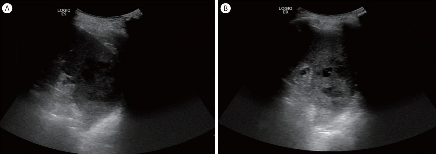
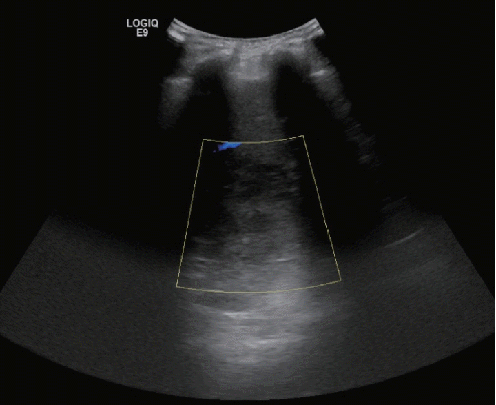
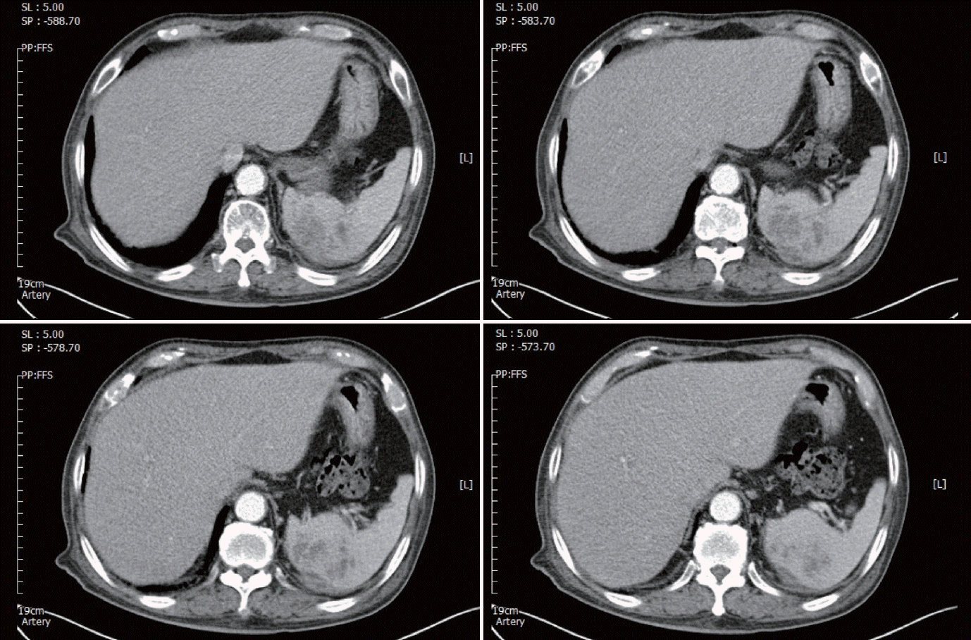
4개월 전부터 폐결핵을 치료하였으나 꾸준히 투약은 하지 못한 85세 남성이 복부 팽창감으로 내원하였다. 피로감을 호소하였고 열은 없었다. 초음파상 5 cm 크기의 불분명한 경계의 저에코와 혼합에코의 종괴 모양과 내부에 저에코의 낭종 소견이었다(Fig. 1). 조영증강 초음파에서도 병변의 경계와 병변 내부의 사이막(septum)에 조영증강이 일부 관찰되다가 소실되는 양상이었다(Fig. 2). CT상 내부의 낭성 병변을 가지는 저밀도의 종괴 모양의 비장농양이 의심되었다(Fig. 3 and 4). 혈액 검사에서 백혈구는 9,210/μL, 혈색소는 11.4 g/dL, 혈소판은 382,000/μL, CRP 2.31 mg/dL였다. 생화학 검사에서 AST/ALT 25/12 U/L, 알칼리인산분해효소 90 U/L, 총 빌리루빈 0.3 mg/dL였다. 종양표지자와 기생충 혈액 검사는 음성이었다.

(A) Ultrasonography showed an about 5 cm-sized hypoechoic and mixed-echoic mass with unclear borders in the spleen. (B) Ultrasonography showed a mass with internal lower echoic cystic lesions in the spleen.

(A) Contrast enhancement is observed in the margin and the septum at the arterial phase. (B) A slight contrast washout was seen at the delayed phase.
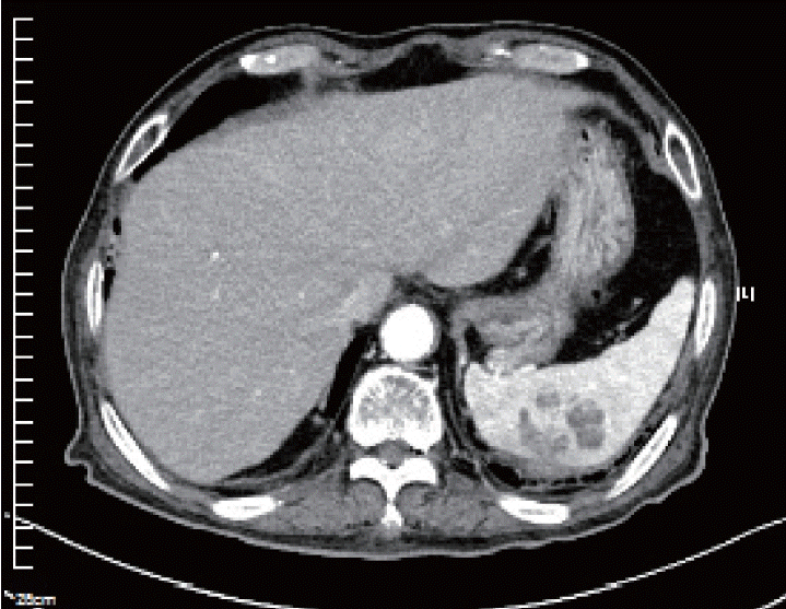
복부 초음파와 CT 소견으로 화농성 비장농양을 의심하였고 입원 후 경험적 항생제를 투약하였다. 입원 중 치료에서 증상 및 혈액 검사 변화가 없어서 항생제를 중단하였고 화농성 비장농양보다 냉농양(cold abscess) 형태의 비장결핵의 가능성을 고려하였다. 결핵약을 이미 복용 중이어서 복약 지도를 하였고 추적 검사를 권유하였다. 환자는 결핵약을 불규칙적으로 복용하였고 5개월 후 추적 초음파(Fig. 5)와 CT (Fig. 6)를 시행하였는데 크기와 모양에 큰 변화가 없었고 증상 악화도 없었다. 결핵약 투약 지속을 권유하였고 외래에서 추적 관찰 중이다.

Follow-up ultrasonography showed an about 5 cm-sized hypoechoic and mixed-echoic mass with inner hypoechoic cysts in the spleen (prone position).
고 찰
비장결핵은 주로 폐결핵에서 혈행성 확산으로 생기고 폐침범 없는 비장결핵도 있을 수 있다[1,2]. 비장결핵은 전신적인 것에서부터 다양한 징후와 증상을 나타낼 수 있다. 피로, 체중 감소, 비장 비대 또는 불명열, 문맥 고혈압 및 비장 파열이 있다[3]. 백혈구수 이상이나 적혈구 증가증이 있을 수 있으나 비특이적이고 초기 영상 방식은 비용 효율적인 복부 초음파이다. 비장 병변은 간비장종대, 농양 등 비특이성 소견 혹은 작은 저에코 결절 또는 더 큰 저에코 종괴가 있을 수 있다[4]. 농양 형태가 가능하며 결핵 치료에도 영상학적 호전이 느린 편이다. 또한 비장결핵은 비장농양 및 다른 비장 질환과 감별이 어렵고 다른 질환으로 오인되기도 한다. 흔하지 않은 비장 병변에서 불명열이나 결핵 치료 중인 환자에서 여러 가지 인자를 고려한 비장결핵 감별진단이 필요하다. 또한 비장결핵이 의심될 경우 치료 및 증상 및 영상학적 추적 검사를 주의 깊게 해야 한다. 복부 초음파는 간편하고 실시간으로 정보를 얻을 수 있으나 민감도가 낮고 검사자에 대한 의존도가 높다. 비장 질환 초기 검사 및 추적 검사에 유용하다. 일부 환자에서 비장 전체가 관찰되지 않는 경우도 있어서 자세 변화 및 복와위 검사가 도움이 될 수도 있고 전체 관찰을 위해 노력해야 한다. 체위 변화 및 압박법으로도 관찰되지 않는 비장 및 복부 질환의 경우 다른 복부 영상 검사의 장단점을 알고 추가 검사도 충분히 고려하여야 한다.


