경과 관찰 중 악성 종양의 초음파 소견을 보이는 갑상선 낭종
Thyroid Cystic Nodules May Have Ultrasonographic Features Mimicking Malignancy during Interval Changes
Article information
Trans Abstract
Ultrasonography (US) features of thyroid nodules can be helpful in identifying those most at risk for malignancy; these features include marked hypoechogenicity, microcalcifications, irregular margins, and a taller-than-wide shape (nonparallel orientation). Some cystic nodules change morphologically after simple aspiration and percutaneous ethanol injection treatment. They can show marked hypoechogenicity and irregular margins on the US and be difficult to distinguish from papillary thyroid carcinoma. This article covered the imaging precautions when a malignant nodule is suspected in the US during the clinical course of a thyroid cyst.
서 론
갑상선 초음파 검사는 다른 검사에 비해 방사선의 피폭이 없고, 검사가 단순하고 반복하기 쉬운 초음파 검사 자체의 장점 외에도 갑상선 결절의 악, 양성 감별과 림프절 전이를 진단할 수 있으며, 갑상선암 수술 후 추적 검사에도 유용하여 갑상선 질환의 관리에 있어 청진기와 같은 역할을 하고 있다[1].
2002년 Kim 등[2]은 갑상선 결절의 악성을 시사하는 초음파 소견으로 저에코 음영, 침상(spiculate) 경계, 미세석회화, 가로 보다 세로가 큰(taller than wide) 경우를 제시하였으며, 이들 변수가 갑상선 결절의 악성과 양성을 감별하는 민감도 93.8%, 특이도 66%를 보고하였다. 이후 2016년 미국갑상선학회와 대한갑상선영상의학회에서 갑상선암 위험도를 초음파 소견으로 분류하여 세침흡인 검사의 기준까지 제시하였다[3,4]. 대한갑상선영상의학회의 Korean Thyroid Imaging Reporting and Data System (K-TIRADS)은 갑상선 결절의 내부 성분, 에코, 세 가지 암 의심 소견(미세석회화, 불규칙 경계, 비평행 방향성)을 기준으로 높은 의심(K-TIRADS 5) (Fig. 1), 중간 의심(K-TIRADS 4), 낮은 의심(K-TIRADS 3), 양성(K-TIRADS 2) 결절로 나누었고, 결절이 없는 경우(K-TIRADS 1)로 분류하였으며, 현재 임상에서 유용하게 이용되고 있다[4]. 초음파 검사 소견은 악성과 유사한 소견을 보이나 실제는 양성 결절인 경우가 있어 임상에서 주의를 요하는 경우를 자주 경험을 한다.
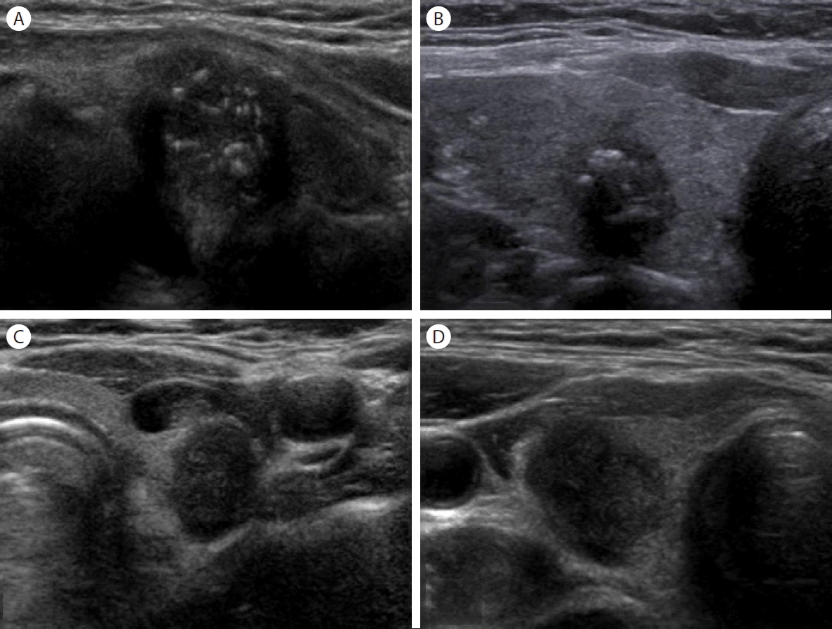
K-TIRADS 5 nodules. Solid hypoechoic nodule with microcalcifications (A), solid hypoechoic nodule with multiple macrocalcifications (B), solid hypoechoic nodule with non-parallel orientation (C), and solid hypoechoic nodule with irregular margin (D). K-TIRADS, Korean Thyroid Imaging Reporting and Data System.
갑상선 낭종은 전체 갑상선 결절의 6-37% 정도로 흔한 양성 종양으로, 초음파 검사에서 한층의 얇은 피막과 내부에 무에코 또는 콜로이드를 함유한 원형 또는 타원형의 결절로 후방 에코 증가가 관찰된다[5]. 갑상선 낭종은 저절로 소실되는 경우도 있으나, 커져 압박 증상을 보이거나 미용상 문제가 있을 경우 단순 흡인으로 낭액을 제거하거나 경피적 에탄올 절제술을 시행하기도 한다. 경피적 에탄올 절제술은 재발성 갑상선 낭성 결절의 치료에서 최대 85-98.5%의 부피 감소 효과를 보이는 유용한 치료 방법으로, 특히 낭성 부분이 90% 이상인 갑상선 결절에서 효과적인 것으로 알려져 있다[5].
이전 갑상선 낭종의 치료로 단순 흡인 시술을 받았거나, 경피적 에탄올 절제술을 받은 경우 갑상선 초음파 검사에서 악성 결절이 의심되는 소견을 보이는 경우가 보고되어 있다[6-8].
본고에서는 갑상선 낭종의 임상 경과 중 초음파 소견에서 악성 결절이 의심될 때 영상학적 주의점을 고찰해 보고자 한다.
본 론
갑상선 낭종은 흔한 갑상선 양성 종양으로 초음파 검사로 쉽게 진단이 가능하다. 그러나 갑상선 낭종 중 단순 흡인 시술이나 경피적 에탄올 절제술을 시행한 경우 시간이 경과한 후에 초음파 검사에서 결절의 크기가 작아지고, 심한 저에코 음영을 보이는 경우 갑상선 암으로 오인을 할 수 있다.
증례 1
60세 여자가 개인의원에서 건강검진 결과 갑상선 초음파 검사에서 악성 결절이 의심되는 병변이 있다고 조직 검사를 위해 내원하였다. 병력 청취에서 환자는 3년 전 본원에서 재발되는 갑상선 낭종으로 경피적 에탄올 절제술을 시행한 적이 있어 이전 초음파를 비교해 보았다. 내원 15개월 전 갑상선 좌엽에 2.60 × 1.92 cm 크기의 낭종이 0.4 cm 크기의 저에코 음영을 동반한 종양으로 변해 있었다(Fig. 2).
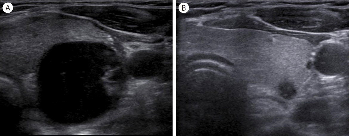
A 60-year-old woman with a cystic thyroid nodule in the left lobe. Transverse ultrasonography (US) before percutaneous ethanol injection therapy (PEIT) shows a large thyroid nodule with a 90% cystic portion with a mural nodule (A). Transverse US obtained 15 months after PEIT shows a marked decrease in nodule size and hypoechogenicity, microcalcification and blurred margins (B).
증례 2
68세 남자가 갑상선 낭종 천자 후 28개월 후 추적 관찰을 위해 내원하였다. 내원 당시 시행한 초음파 검사에서 갑상선 좌엽에 1.01 × 1.10 cm 크기의 불규칙한 변연부를 가진 저에코 음영의 결절이 관찰되었다. 내원 28개월 전 시행한 초음파 검사에서는 2.05 × 1.62 cm 크기의 낭종이 관찰되었다(Fig. 3).
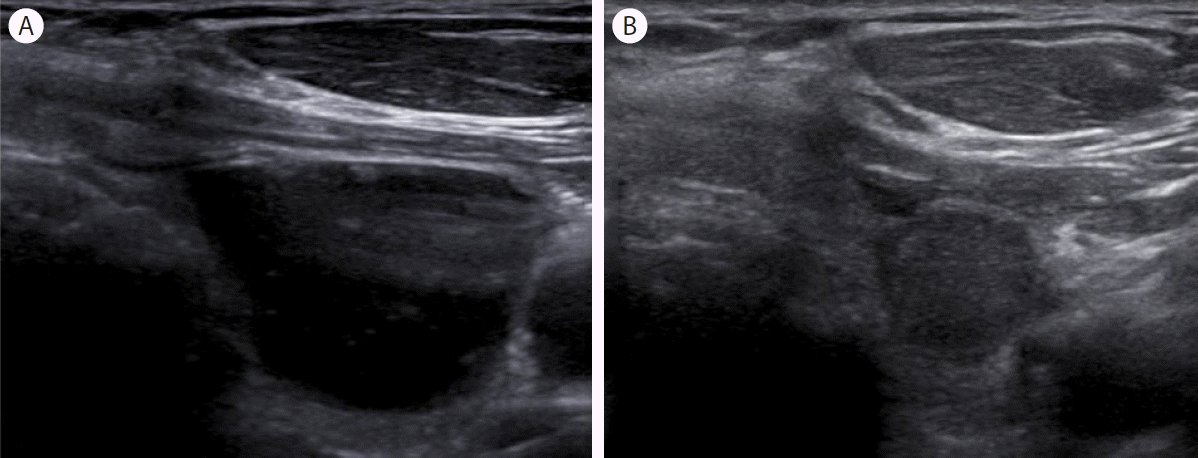
A 68-year-old man with a cystic thyroid nodule in the left lobe. Transverse ultrasonography (US) image before aspiration shows a large cystic nodule with colloid material (A). Transverse US obtained after 28 months shows a decrease in nodule size, along with hypoechogenicity, microcalcification, and irregular margins (B).
증례 3
63세 남자가 갑상선 낭종 천자 후 23개월 후 추적 관찰을 위해 내원하였다. 내원 당시 시행한 초음파 검사에서 갑상선 우엽에 0.47 × 0.48 cm 크기의 미세석회화, 불규칙한 변연부를 가진 저에코 음영의 결절이 관찰되었다. 내원 23개월 전 시행한 초음파 검사에서는 갑상선 우엽에 1.66 × 1.41 cm 크기의 낭종 우세 결절이 관찰되었다(Fig. 4).
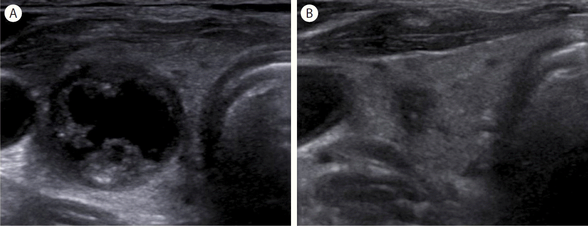
A 63-year-old man with a predominant cystic thyroid nodule in the right lobe. Transverse ultrasonography (US) before aspiration shows a 1.66 × 1.41 cm sized predominant cystic nodule (A). Transverse US performed after 23 months shows a 0.47 × 0.48 cm sized nodule with hypoechogenicity, microcalcification, and blurred margins (B).
증례 4
75세 여자가 갑상선 낭종 천자를 시행받고 25개월 후 추적 관찰을 위해 내원하였다. 내원 당시 시행한 초음파 검사에서 갑상선 우엽에 0.96 × 1.11 cm 크기의 저에코 음영의 불규칙한 변연부와 갑상선외 침범 소견을 보이는 결절이 관찰되어 세침흡인세포 검사를 시행하였으며, 양성 결절로 진단되었다. 환자의 내원 25개월 전 시행한 초음파 검사에서는 갑상선 우엽에 1.60 × 1.32 cm 크기의 낭종 우세 결절이 관찰되었다(Fig. 5).

A 75-year-old woman with a cystic thyroid nodule in the right lobe. Transverse ultrasonography (US) before aspiration shows a 1.60 × 1.32 cm-sized predominant cystic nodule (A). Transverse US performed after 25 months shows a 0.96 × 1.11 cm sized nodule with hypoechogenicity, irregular margin, and extrathyroidal extension (B).
증례 5
26세 여자 환자가 갑상선 낭종 천자 후 경과 관찰을 위해 내원하였다. 내원 28개월 전 처음 방문하였을 때 초음파 검사에서 갑상선 우엽에 2.42 × 1.85 cm 크기의 낭종 우세 양성 종양 소견을 보였으며, 낭종 흡인 시술 7개월 후인 내원 21개월 전 추적 검사에서 갑상선 우엽에 1.01 × 1.10 cm 크기의 저에코 음영과 불규칙한 변연부를 보이는 종양이 관찰되었다. 마지막 내원 시에는 결절의 크기가 0.27 × 0.20 cm 크기로 현저히 줄어져 있었고, 저에코 음영과 불규칙한 변연부 소견을 보였다(Fig. 6).
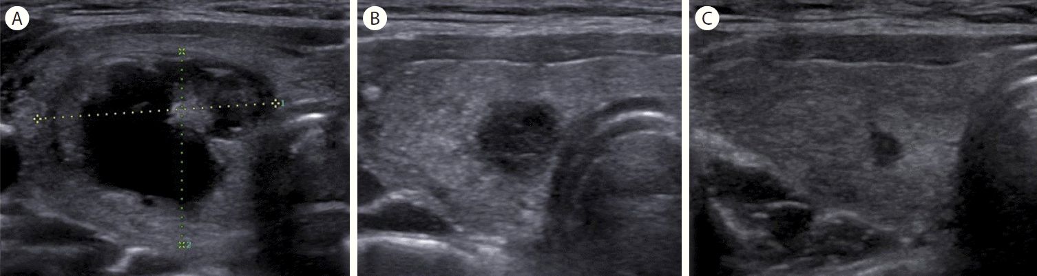
A 26-year-old woman with a predominant cystic thyroid nodule in the right lobe. A transverse ultrasonography (US) image obtained before aspiration shows a 2.42 × 1.85 cm-sized predominant cystic nodule (A). A transverse US image obtained after 7 months shows a 1.01 × 1.10 cm sized nodule with hypoechogenicity and irregular margin (B). Transverse US performed after 28 months shows a large decrease in size, along with hypoechogenicity and irregular margin (C).
Koo 등[7]은 낭종 천자 후 초음파 검사에서 악성 결절로 보이는 31개의 결절에 대해 14예에서는 추가적인 세침흡인세포 검사를 시행하였으며, 모두 양성 결절로 진단되었고 한다. 본 증례 4의 경우에도 추적 초음파 검사 소견상 갑상선 결절의 모양이 악성 소견을 보여 세침흡인세포 검사를 시행하였고 양성 결절로 진단되었다.
결 론
갑상선 낭종의 경우 시간이 지남에 따라 초음파 검사에서 모양이 변하여 악성 결절로 오진될 수도 있으므로 임상적으로 유의해야 한다. 임상에서 갑상선 초음파 검사상 악성을 시사하는 소견을 보이는 결절이 있을 경우에는 반드시 이전에 갑상선 낭종에 대한 흡인 시술이나 경피적 에탄올 절제술을 시행한 병력 등을 문진이나 이전에 촬영한 초음파 영상을 통하여 확인하는 것이 중요하다.